Husseing and Retamal, December 2005
Introduction
It is becoming increasingly apparent that AAGP® has strong cell preservation qualities. With this in mind a series of experiments mimicking cell stress conditions (pH, oxidative stress, low temperatures and time) were conducted to challenge both human adult and neonatal fibroblasts exposed to AAGP® in comparison to human adult and neonatal fibroblasts not exposed to AAGP®. This was done with the use of AAGP® as a anti- aging cosmetic, protecting skin from the harsh environment, in mind. The first of these experiments is outlined below and aimed to elucidate the protective effects of AAGP® at low temperatures (-3°C and 3°C)
Method
Results
The ability of AAGP® to protect against temperature induced cell death can be seen as early as 1hour into the experiment. By 34 hours cells exposed to temperatures of -3 or 3°C were at only 20% viability when in the presence of no AAGP®. However, at both temperatures cells exposed to 10 or 15mg/ml AAGP® maintained viability percentages of above 80% (96% for 10 and 15mg/ml AAGP® exposed cells at -3°C).
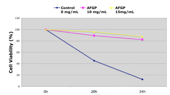
Figure 1: Effects of AAGP™ on the survival of human neonatal fibroblasts at 3°C.
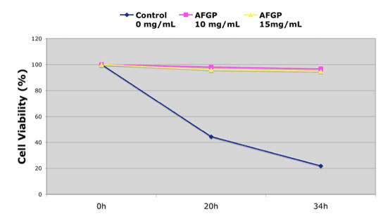
Figure 2: Effects of AAGP™ on the survival of human neonatal fibroblasts at -3°C.
Conclusion
These results conclude that AAGP® is able to protect cells against low-temperature induced cell death for up to 34 hours. The mechanism by which this occurs is presently unknown.